1 of 2 product images
Molecular Products
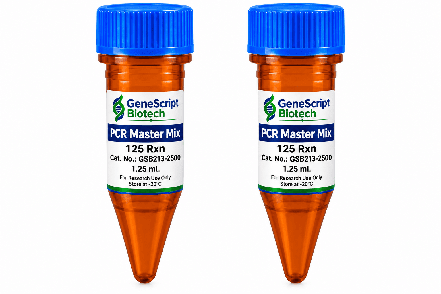
PCR Master Mix 250 Rxn
This PCR Master Mix also called amaR PCR Master Mix is light sensitive and should be stored and protected from light.
- Catalog no.
- GSB213-2500
- Subcategory
- amaR PCR Master Mix 250 Rxn
Ask about pricing, availability, configuration or technical fit. No account required.
Description
The PCR Master Mix is a ready-to-use PCR reaction mixture. Simply add primers, template, and water, the reagent will execute primer extensions and other molecular biology applications. The PCR Master Mix is a pre-mixed solution containing Taq DNA polymerase, PCR buffer, dNTPs, gel loading dyes, and fluorescence dye. The PCR Master Mix which contains the Taq DNA polymerase, is purified from the E. coli., and expressing the Thermus aquaticus DNA polymerase gene. This enzyme has a 5' → 3' DNA polymerase and the 5' → 3' exonuclease activity but lacks the 3' → 5' exonuclease activity. The PCR Master Mix, which contains the fluorescence dye, is directly detected on BLooK LED transilluminator or UV epi-illuminator after the DNA electrophoresis. The PCR Master Mix contents red tracking dyes, provide a safe, non-toxic and non-mutagenic alternative to ethidium bromide for instantaneous band visualization, that are environmentally friendly containing no hazardous chemicals. The tracking dyes that run at 10 bp on a 1% agarose gel. The PCR Master Mix mixture is supplied at the 2X concentration to allow approximately 50% of the final reaction volume to be used for the addition of primer and template solutions. The reagents are provided with the sufficient amplification reactions of 20 μl each.
Features
- No post-staining processing of DNA required.
- No need to prepare PCR Reagents.
- Direct loading onto your agarose gel for analysis.
- Sensitivity – High degree of sensitivity as the ethium bromide.
- Speed – No destaining requirement.
- Compatibility – Use the blue light or UV to detect the signal.
Tracking dye
- Amaranth
Product data
Technical specifications
| Protocol | Standard PCR with PCR Master Mix | |
|---|---|---|
| 1. For each 20 μl reaction, assemble the following components in a 0.2 ml PCR tube on ice just prior to use: | ||
| ======= | Volume_ | Final Concentration |
| PCR Master Mix | __10 (μl) | 1X |
| Forward primer, 5~10 μM | Variable | 0.1-0.2 μM |
| Reverse primer, 5~10 μM | Variable | 0.1-0.2 μM |
| DNA template | Variable | 4 pg~500 ng |
| Add ddH2O to | 20 (μl) | |
| 2. Mix gently. If necessary, centrifuge briefly, Cap tubes and place in the thermal cycler. | ||
| 3. Process in the thermal cycler for 25~35 cycles as follows: | ||
| Initial Denaturation | 2~5 minutes at 94°C | |
| Denaturation | 20~40 seconds at 94°C | |
| Annealing | 1 minute at the proper annealing temperature | |
| Extension | 2 minutes at 72°C, (Repeat 30 Cycles Denaturation, Annealing & Extension) | |
| Final Extension | 5 minutes at 72°C | |
| All products are research use only. | ||
Note
Optimal conditions for amplification will vary depending on the primers and thermal cycler used. It may be necessary to optimize the system for individual primers, template, and thermal cycler.
- After the PCR reaction, DNA electrophoresis will detect the PCR product.
- Use the UV or blue-light transilluminator or UV epi-illuminator to photograph the gel. Note: When the DNA concentration is less than 4 pg, it may cause the migratory shift when performing the electrophoresis. To remedy this observation, we recommend to conduct the following steps (please refer to the experimental procedures), or use the PCR Clean-Up & Gel Extraction Kit (see GSB006-0100) to remove the flrorescence dye prior to post-staining with the Novel Green (GSB002-0500) or Novel Green plus (GSB003-0500) again for restoring the DNA molecular weight in the original position.
Removal of Fluorescence Dye
- Immerse the PCR product containing the fluorescence dye into the 100 mM NaCl and add 2.5 volumes of absolute or 95% ethanol.
- Incubate on ice for 20 minutes.
- Centrifuge the mixture at 4°C for at least 10 minutes.
- Remove the suspension of ethanol and wash the pellet with 1ml of 70% ethanol.
- Dry the residual ethanol and resuspend the double-stranded DNA in the TE.
Explore more